About this Event
From Spatial Transcriptomics to Quantitative Protein Phenotypes: Imaging Mass Cytometry in Cancer, Neuroscience and Translational Research
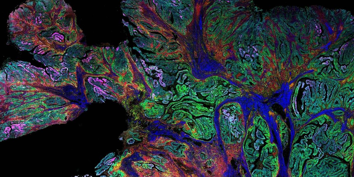
Imaging Mass Cytometry™ (IMC™) technology enables high-dimensional per-cell protein quantitation in intact tissue, providing a powerful framework for studying cellular phenotypes and tissue architecture in complex biological systems. By measuring dozens of proteins simultaneously at single-cell resolution, IMC workflows allow researchers to map tumor–immune interactions, define immune and stromal cell states, and quantify clinically relevant biomarkers directly within the tumor microenvironment and other tissues. These capabilities are increasingly important across immuno-oncology, neuroscience and translational research. IMC technology also complements emerging spatial transcriptomic platforms such as Xenium by enabling direct validation and quantitation of protein expression in the same cells and tissue context where RNA signatures were measured.
This seminar will be held in Dorothy M. Davis Heart and Lung Research Institute (DHLRI) room 165.
Event Venue & Nearby Stays
Dorothy M. Davis Heart & Lung Research Institute, 473 West 12th Avenue, Columbus, United States
USD 0.00